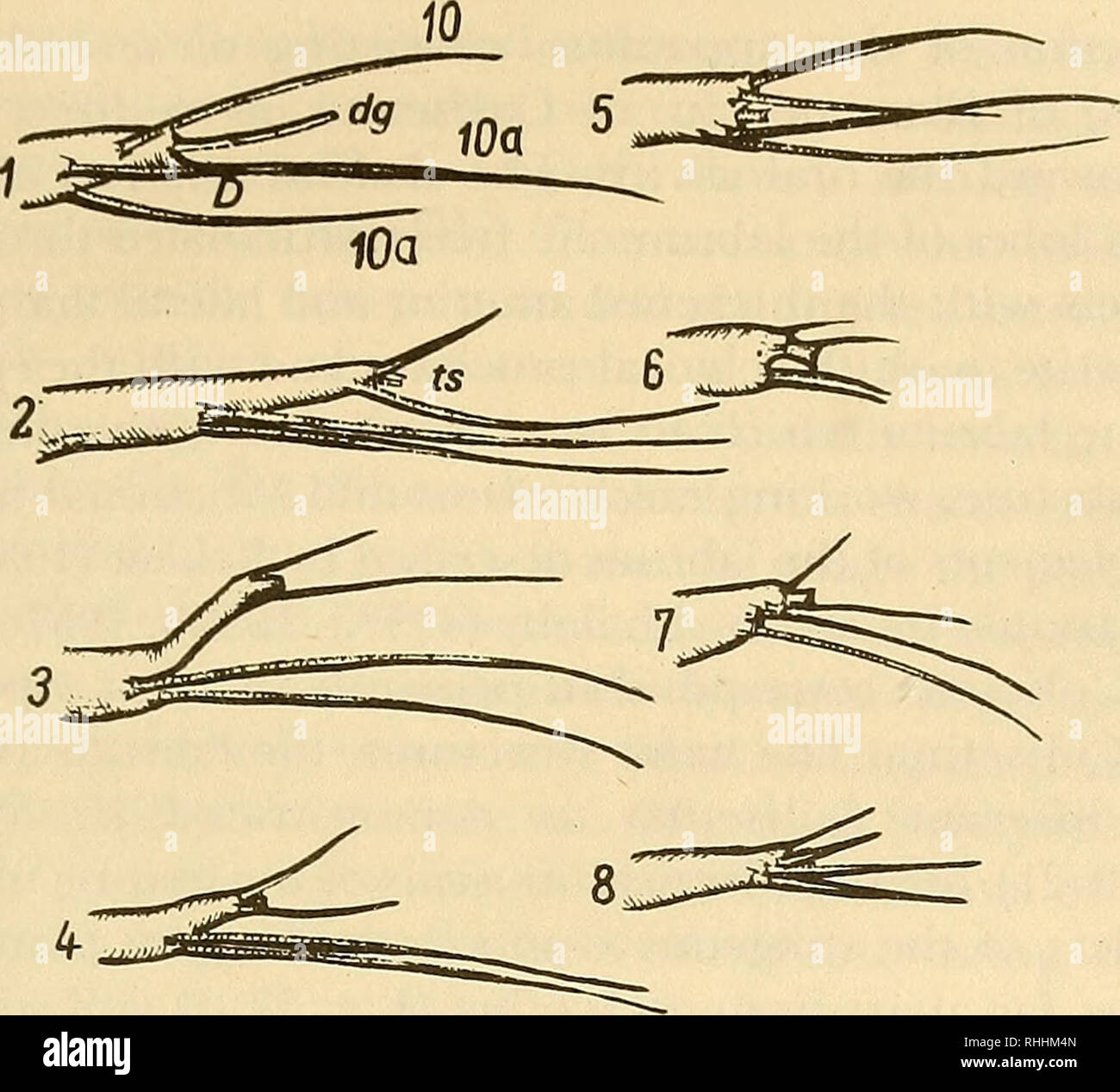
. Blood-sucking mosquitoes of the subtribe Culisetina (Diptera, Culicidae) in world fauna. Mosquitoes. 63. Fig. 35. Structure and types (1-8) of apical antennal appendages. -Dolabriform appendage; dg—digitate appendage; ts—terminal spine; 10—terminal spinule (tsp); 10a—subterminal spinule (stsp). have been conducted on the structure and functioning of the mouth apparatus of Anopheles larvae and certain species of Culex and Aedes. In a special publication describing a detailed study of the mouth parts of Culiseta incidens larvae (Cook, 1949), there is an excellent description of the muscle syst
Image details
Contributor:
Library Book Collection / Alamy Stock PhotoImage ID:
RHHM4NFile size:
7.1 MB (263.8 KB Compressed download)Releases:
Model - no | Property - noDo I need a release?Dimensions:
1660 x 1504 px | 28.1 x 25.5 cm | 11.1 x 10 inches | 150dpiMore information:
This image is a public domain image, which means either that copyright has expired in the image or the copyright holder has waived their copyright. Alamy charges you a fee for access to the high resolution copy of the image.
This image could have imperfections as it’s either historical or reportage.
. Blood-sucking mosquitoes of the subtribe Culisetina (Diptera, Culicidae) in world fauna. Mosquitoes. 63. Fig. 35. Structure and types (1-8) of apical antennal appendages. -Dolabriform appendage; dg—digitate appendage; ts—terminal spine; 10—terminal spinule (tsp); 10a—subterminal spinule (stsp). have been conducted on the structure and functioning of the mouth apparatus of Anopheles larvae and certain species of Culex and Aedes. In a special publication describing a detailed study of the mouth parts of Culiseta incidens larvae (Cook, 1949), there is an excellent description of the muscle system of the labrum, labium 53 and pharynx but morphological study of the maxillae is absent. It is therefore necessary to describe in some detail the individual mouth parts of Culisetina larvae since among Culisetina we find distinct differences of larval feeding such as filter feeding and typical scrap- ing of vegetation. Our description may not, however, be complete. Although the mouth parts of the various larvae differ signifi- cantly in structure depending on the nature of feeding, feeding by Culisetina larvae is not strictly confined to one method. Typical filter feeders (subgenus Culicella) are capable of scraping vegetation as typical periphy to phages such as Allotheobaldia longiareolata are capable of filter feeding on plankton. Consequently, even the mor- phological structures of these larvae preserve versatility to some extent which is variously manifest in the different members. The labrum is in the form of a complex system of freely moving setae and filaments forming a single, unpaired median and two lateral lobes or flabellae (or tufts; flabellum of foreign authors). The. Please note that these images are extracted from scanned page images that may have been digitally enhanced for readability - coloration and appearance of these illustrations may not perfectly resemble the original work.. Maslov, A. V; Ward, Ronald A. Washington : Smithsonian Institution Lib