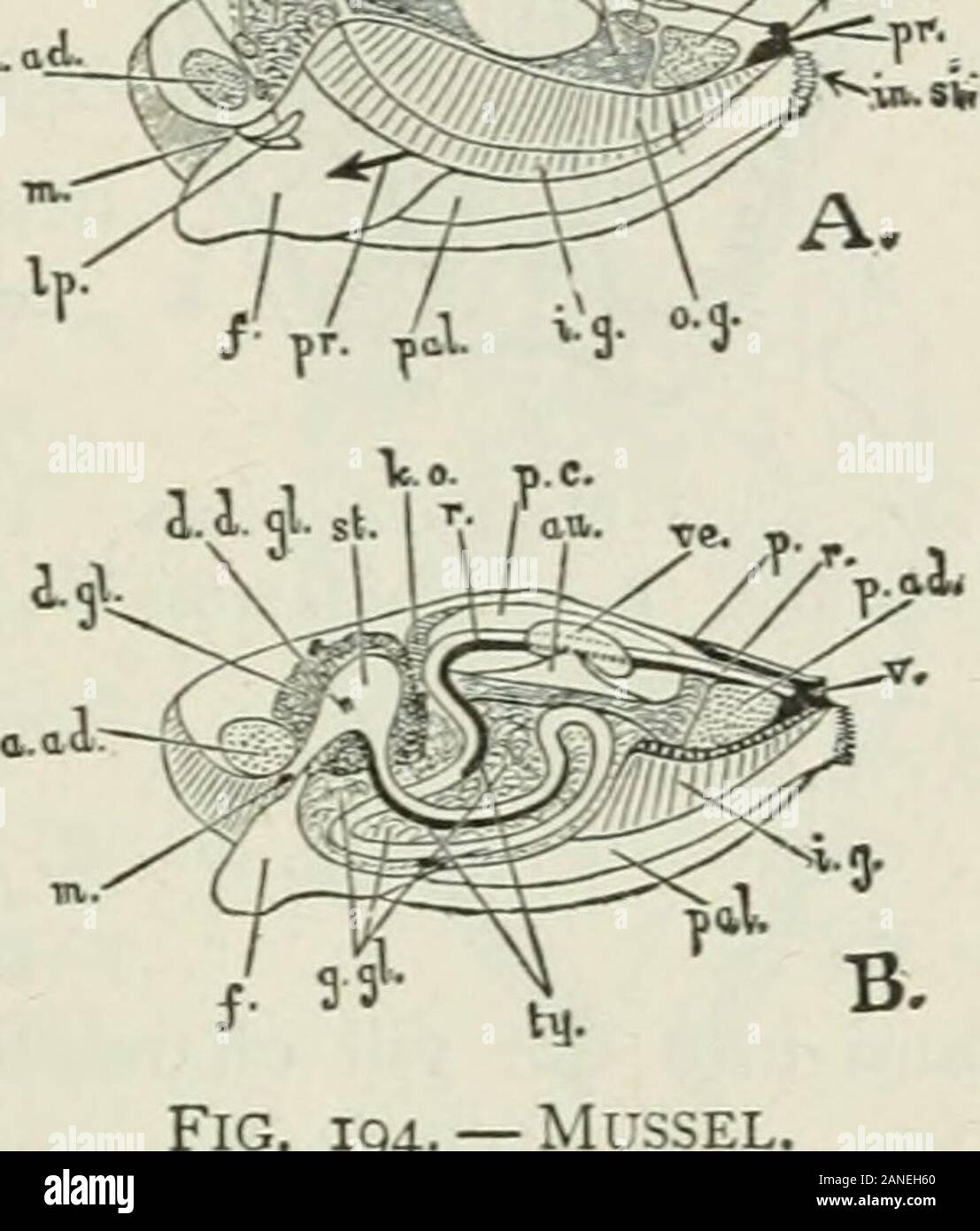
Beginners' zoology . Fig. 193. — Anatomy of Mussel. (Beddard.) MOLLUSCS lOI chamber for the gills is made by the joining of the mantleflaps below, along the ventral line. The mantle edges areseparated at two places, leaving openings called cxJialejitand inhaleJit sipJions. Fresh water with its oxygen, propelled by cilia at theopening and on the gills, enters through the lower orinhalent siphon, passes between the gills, and goes to anupper passage, leaving the gill chamber by a slit whichseparates the gills from the foot. ?X^. For this passage, see arrow(Fig, 194). The movement ofthe water is

Image details
Contributor:
The Reading Room / Alamy Stock PhotoImage ID:
2ANEH60File size:
7.2 MB (218.7 KB Compressed download)Releases:
Model - no | Property - noDo I need a release?Dimensions:
1459 x 1713 px | 24.7 x 29 cm | 9.7 x 11.4 inches | 150dpiMore information:
This image is a public domain image, which means either that copyright has expired in the image or the copyright holder has waived their copyright. Alamy charges you a fee for access to the high resolution copy of the image.
This image could have imperfections as it’s either historical or reportage.
Beginners' zoology . Fig. 193. — Anatomy of Mussel. (Beddard.) MOLLUSCS lOI chamber for the gills is made by the joining of the mantleflaps below, along the ventral line. The mantle edges areseparated at two places, leaving openings called cxJialejitand inhaleJit sipJions. Fresh water with its oxygen, propelled by cilia at theopening and on the gills, enters through the lower orinhalent siphon, passes between the gills, and goes to anupper passage, leaving the gill chamber by a slit whichseparates the gills from the foot. ?X^. For this passage, see arrow(Fig, 194). The movement ofthe water is opposite to the waythe arrow points. After goingupward and backward, the wateremerges by the exhalent siphon.The gills originally consisted ofa great number of filaments.These are now united, , ^but notcompletely so, and the gills stillhave a perforated or latticestructure. Thus they present alarge surface for absorbing oxy-gen from the water. The mouth is in front of the foot, between it and theanterior adductor muscle (Fig. 194). On each side of themouth are the labial palps, which are lateral lips (Fig. 195).They have ciHa which convey the food to the mouth afterthe inhalent siphon has sent food beyond the gill chamberand near to the mouth. Thus both food and oxygen enterat the inhalent siphon. The foot is in the position of alower lip, and if regarded as a greatly extended lower lip, the animal may be said to have what is to us the absurdhabit of using its lower lip as a foot. The foot is some- FiG. 194.- A, left she